
Muscle Quality and Heart Health in Dialysis Patients: CT Study
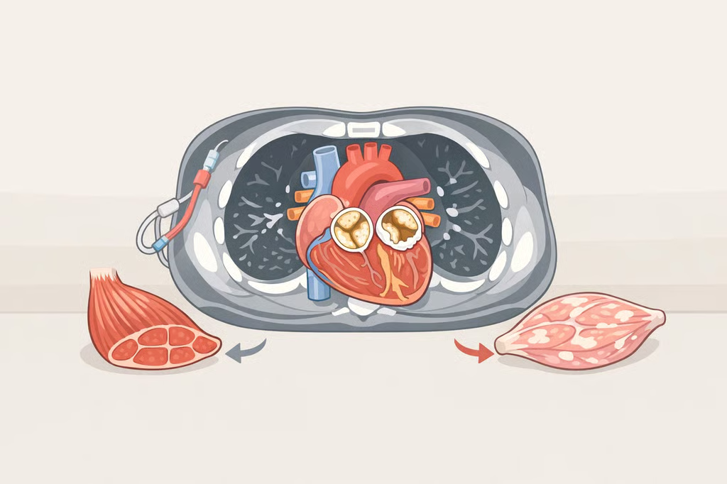
Dialysis patients with lower-density skeletal muscle have higher odds of calcified heart valves than patients with denser muscle, even though muscle mass itself showed no such association, according to a study of four dialysis centers in China published last year in Frontiers in Physiology. The finding adds specific numbers to an emerging question around muscle quality and heart health in dialysis patients, a population in which more than a third of scanned patients, 36.5%, already had detectable calcification on their heart valves, the study found.
Researchers analyzed chest CT scans and echocardiograms from dialysis patients across the four centers. Each one-standard-deviation drop in skeletal muscle density raised the adjusted odds of valve calcification by 20%, according to the study. Patients in the lowest density quartile had 49% higher odds of valve calcification than those in the highest quartile, after accounting for other health factors, the research showed.
Skeletal muscle index, a separate CT-based measure of how much muscle a patient has rather than what it's made of, showed no independent association with valve calcification once researchers adjusted for other variables, the study reported. The finding suggests muscle composition may carry information that differs from muscle quantity, though the study did not test whether measuring muscle density changes clinical decision-making.
Skeletal muscle density and cardiac valve calcification
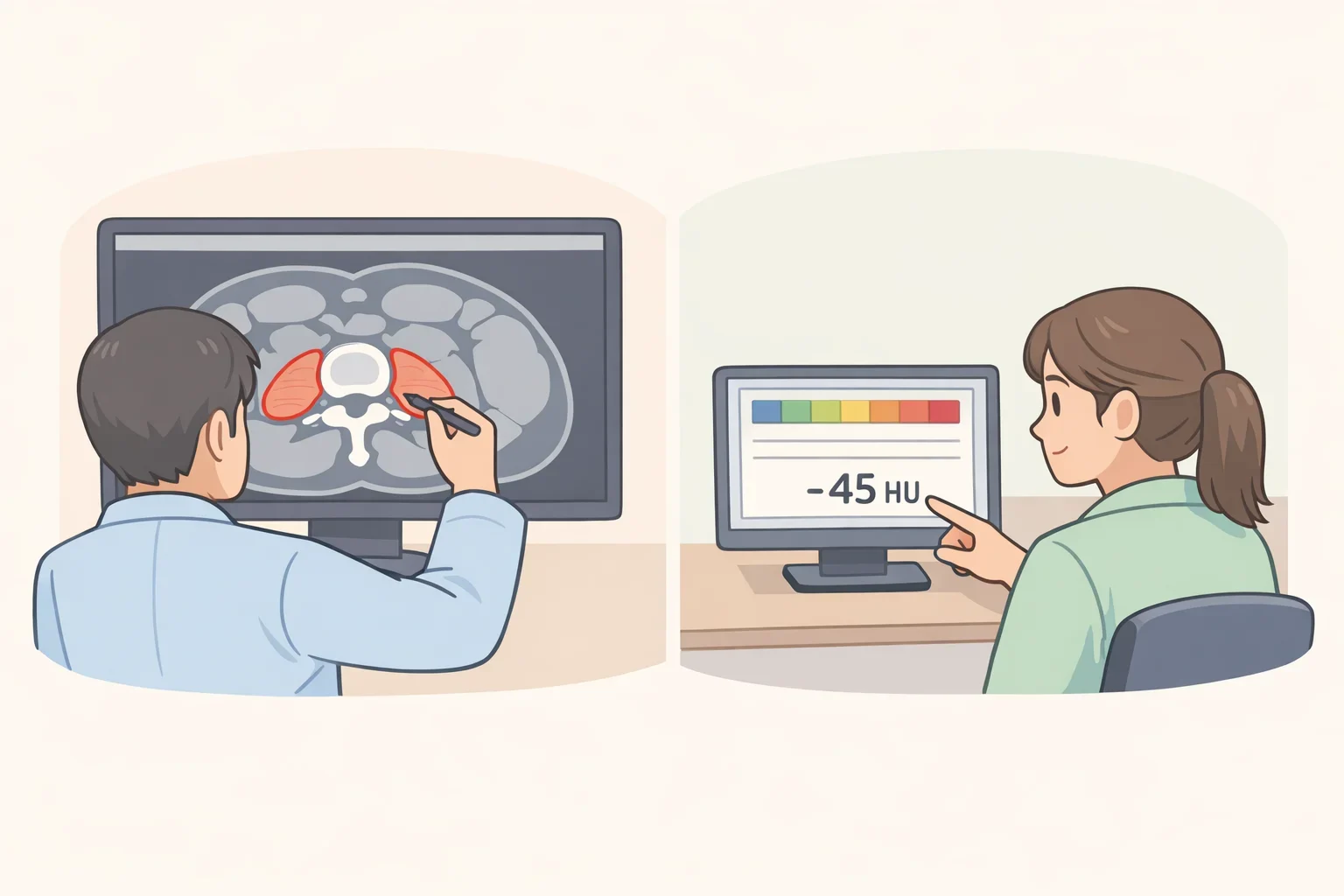
Researchers calculated skeletal muscle density (SMD) from the average CT radiation attenuation across the muscle's cross-sectional area at the L1 vertebra, a measurement that reflects muscle quality rather than muscle size, the study explained. Low muscle density in dialysis patients describes what the tissue is made of, since more fat infiltrating the muscle lowers the attenuation reading, not how large the muscle is.
The scans used weren't ordered to check muscle composition in the first place. They came from chest CT taken for other clinical reasons and later analyzed alongside echocardiogram results for both muscle density and valve calcification, the researchers noted.
The study's authors proposed a cutoff of 31.16 Hounsfield units to flag higher-risk patients. That threshold correctly identified 53.7% of true cases and correctly ruled out 68.6% of patients without valve calcification, the study found. A sensitivity of 53.7% means the cutoff would miss close to half of patients who actually have valve calcification; the study does not establish this cutoff as a stand-alone screening test.
Three studies, three different heart problems
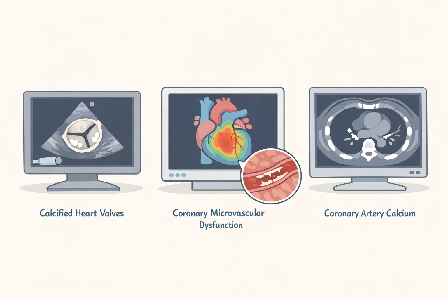
The dialysis study measured calcification on heart valves. A separate cohort, published last year, tracked coronary microvascular dysfunction (CMD), a problem with blood flow through the heart's small vessels, alongside heart attacks, heart-failure hospitalizations, and death. A third analysis, from the Multi-Ethnic Study of Atherosclerosis (MESA), measured calcium buildup in the coronary arteries themselves. These are three different structures and disease processes, not interchangeable stand-ins for "heart health."
That coronary cohort followed 669 patients referred for cardiac stress testing with positron emission tomography, all showing normal perfusion and preserved left-ventricular ejection fraction at the time of testing, according to the study. None were dialysis patients. Researchers tracked outcomes over a median of six years and used CT scans originally captured for PET attenuation correction, a routine technical step in that imaging process, to measure muscle and fat at the 12th thoracic vertebra.
Patients with more intermuscular adipose tissue (IMAT), the CT-derived measure of fat infiltrated into muscle, faced a higher risk of major adverse cardiovascular events (MACE) during follow-up, independent of body mass index. The hazard ratio was 1.53 for every 10-square-centimeter increase in IMAT, the researchers found. Patients with both reduced coronary blood flow and a higher fatty-muscle fraction carried the highest MACE risk of any group in the study, a combination the authors described as a novel at-risk cardiometabolic phenotype, according to the research.
That same cohort complicates any simple story about muscle quality replacing muscle mass. Larger skeletal muscle area was independently protective, lowering MACE risk by 11% for every 10-square-centimeter increase, even as higher IMAT raised risk in the same statistical model, the study reported. In the dialysis cohort, by contrast, muscle mass showed no independent link to valve calcification at all. Muscle quality may add information beyond size in some contexts, but these two studies don't agree on whether size stops mattering.
A third study complicates things further. In MESA's larger, community-based sample of 1,974 adults free of coronary heart disease, abdominal muscle area and density were not independently linked to coronary artery calcification (CAC) once other factors were accounted for, the researchers found. Visceral-fat area and density, rather than abdominal muscle measures, were associated with the presence of CAC after adjustment in that population, according to the study.
Whether muscle quality shows up as meaningful depends on which heart structure, which population, and which measurement method a study uses. Taken together, the three studies do not show that one muscle measure predicts every form of cardiovascular disease.
What the studies can't answer yet
None of these three studies can establish cause and effect. Each is observational: researchers measured muscle composition and cardiovascular findings in existing patient groups rather than testing whether changing muscle density changes outcomes.
The dialysis study is cross-sectional, meaning muscle density and valve calcification were measured at a single point in time for each patient, according to the research. That design can show an association but can't establish which came first, or whether treating low muscle density would change valve outcomes.
The dialysis findings also come from four centers in one country, and the proposed density cutoff had modest sensitivity and specificity, as noted above. Whether the same threshold or association would hold in other dialysis populations, with different demographics, dialysis approaches, or scanning protocols, has not been tested.
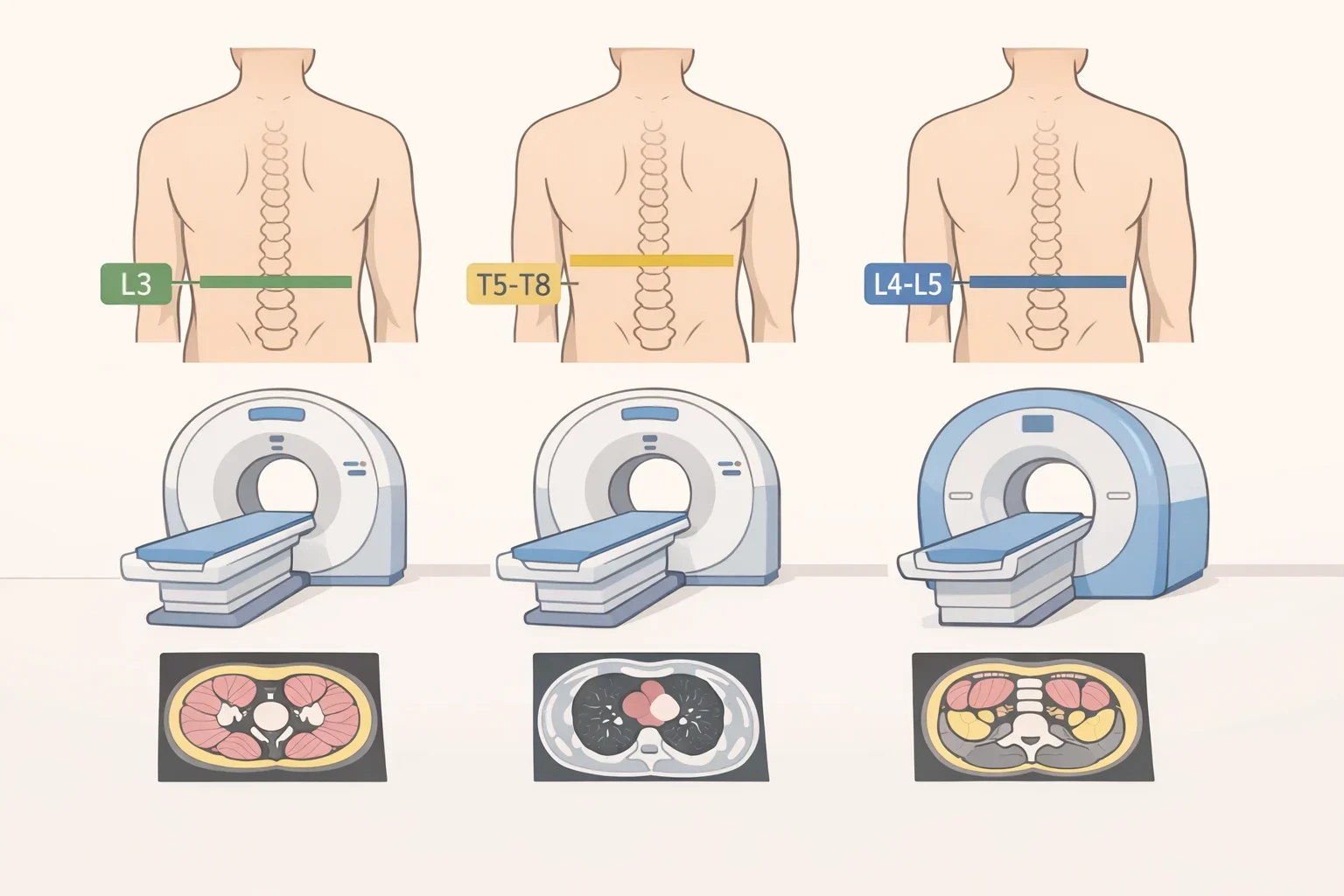
Measurement methods differ across the research, too. The dialysis study measured SMD at the L1 vertebra using chest CT. The coronary cohort measured IMAT at the T12 vertebra using PET attenuation-correction CT. MESA measured abdominal muscle area and density across the L2 to L5 vertebrae using dedicated abdominal CT. There is no single agreed-upon scan site or Hounsfield-unit threshold for "muscle quality" across this research, which makes comparing results between studies, or applying one study's cutoff to a different population, difficult.
The coronary cohort, while it followed patients for a median of six years and adjusted for BMI, drew from people already referred for coronary evaluation, not from dialysis patients or the general population, the study noted. Its findings about IMAT, muscle area, and MACE risk may not carry over to people undergoing dialysis, whose muscle loss and cardiovascular risk factors differ substantially from that group.
None of the three studies tested whether increasing muscle density, through resistance training, nutrition changes, or medical treatment, changes valve calcification, cardiovascular events, or survival. They establish associations at a single point in time or over follow-up, not treatment targets.
What this means right now

These findings do not establish a reason to order a CT scan solely to measure muscle density. All three studies used scans that were already performed, or scan data already captured, for other clinical purposes.
For patients managing dialysis or known cardiovascular risk factors who already undergo CT imaging or echocardiography as part of their care, muscle-density findings are a research signal a nephrologist or cardiologist may eventually weigh alongside standard monitoring. They are not a replacement for blood pressure checks, kidney function labs, lipid panels, or established cardiac imaging. Questions about how kidney disease or cardiovascular risk relates to muscle health are best raised with a nephrologist, cardiologist, or registered dietitian familiar with the individual case.
In this dialysis cohort, low skeletal muscle density, not low muscle mass, was independently associated with cardiac valve calcification, with odds rising as density quartiles declined. That association has not yet been validated as a screening tool or tied to a treatment that changes outcomes, so it belongs alongside standard cardiovascular and kidney-disease monitoring rather than in place of it.